| Record Information |
|---|
| Version | 2.0 |
|---|
| Creation Date | 2010-05-12 17:30:02 UTC |
|---|
| Update Date | 2014-12-24 20:26:29 UTC |
|---|
| Accession Number | T3D3750 |
|---|
| Identification |
|---|
| Common Name | Aurovertin B |
|---|
| Class | Small Molecule |
|---|
| Description | Aurovertin B is a mycotoxin and antibiotic produced by the fungus Calcarisporium arbuscula. It is known for its ability to inhibit oxidative phosphorylation. (1, 2) |
|---|
| Compound Type | - Ester
- Ether
- Fungal Toxin
- Mycotoxin
- Natural Compound
- Organic Compound
|
|---|
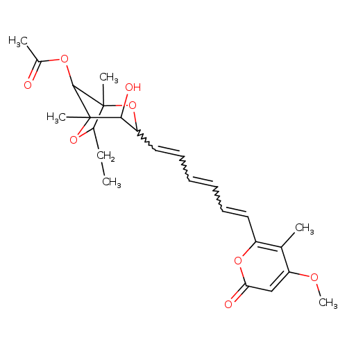
| Chemical Structure | |
|---|
| Synonyms | | Synonym | | aurovertin b | | Aurovertin b | | Aurovertin-B |
|
|---|
| Chemical Formula | C25H32O8 |
|---|
| Average Molecular Mass | 460.517 g/mol |
|---|
| Monoisotopic Mass | 460.210 g/mol |
|---|
| CAS Registry Number | 55350-03-3 |
|---|
| IUPAC Name | 7-ethyl-4-hydroxy-3-[6-(4-methoxy-5-methyl-2-oxo-2H-pyran-6-yl)hexa-1,3,5-trien-1-yl]-1,5-dimethyl-2,6-dioxabicyclo[3.2.1]octan-8-yl acetate |
|---|
| Traditional Name | 7-ethyl-4-hydroxy-3-[6-(4-methoxy-3-methyl-6-oxopyran-2-yl)hexa-1,3,5-trien-1-yl]-1,5-dimethyl-2,6-dioxabicyclo[3.2.1]octan-8-yl acetate |
|---|
| SMILES | CCC1OC2(C)C(OC(C)=O)C1(C)OC(C=CC=CC=CC1=C(C)C(OC)=CC(=O)O1)C2O |
|---|
| InChI Identifier | InChI=1S/C25H32O8/c1-7-20-24(4)23(30-16(3)26)25(5,33-20)22(28)18(32-24)13-11-9-8-10-12-17-15(2)19(29-6)14-21(27)31-17/h8-14,18,20,22-23,28H,7H2,1-6H3/b9-8+,12-10+,13-11+ |
|---|
| InChI Key | InChIKey=QXCOFYWOWZJFEA-YIDQAKLOSA-N |
|---|
| Chemical Taxonomy |
|---|
| Description | belongs to the class of organic compounds known as c-glycosyl compounds. These are glycoside in which a sugar group is bonded through one carbon to another group via a C-glycosidic bond. |
|---|
| Kingdom | Organic compounds |
|---|
| Super Class | Organic oxygen compounds |
|---|
| Class | Organooxygen compounds |
|---|
| Sub Class | Carbohydrates and carbohydrate conjugates |
|---|
| Direct Parent | C-glycosyl compounds |
|---|
| Alternative Parents | |
|---|
| Substituents | - C-glycosyl compound
- 1,4-dioxepane
- Alkyl aryl ether
- Pyranone
- Dioxepane
- Pyran
- Oxane
- Monosaccharide
- Heteroaromatic compound
- Vinylogous ester
- Tetrahydrofuran
- Carboxylic acid ester
- Secondary alcohol
- Lactone
- Oxacycle
- Carboxylic acid derivative
- Organoheterocyclic compound
- Dialkyl ether
- Monocarboxylic acid or derivatives
- Ether
- Alcohol
- Hydrocarbon derivative
- Organic oxide
- Carbonyl group
- Aromatic heteropolycyclic compound
|
|---|
| Molecular Framework | Aromatic heteropolycyclic compounds |
|---|
| External Descriptors | Not Available |
|---|
| Biological Properties |
|---|
| Status | Detected and Not Quantified |
|---|
| Origin | Exogenous |
|---|
| Cellular Locations | |
|---|
| Biofluid Locations | Not Available |
|---|
| Tissue Locations | Not Available |
|---|
| Pathways | Not Available |
|---|
| Applications | Not Available |
|---|
| Biological Roles | Not Available |
|---|
| Chemical Roles | Not Available |
|---|
| Physical Properties |
|---|
| State | Solid |
|---|
| Appearance | White powder. |
|---|
| Experimental Properties | | Property | Value |
|---|
| Melting Point | Not Available | | Boiling Point | Not Available | | Solubility | Not Available | | LogP | Not Available |
|
|---|
| Predicted Properties | |
|---|
| Spectra |
|---|
| Spectra | | Spectrum Type | Description | Splash Key | Deposition Date | View |
|---|
| Predicted LC-MS/MS | Predicted LC-MS/MS Spectrum - 10V, Positive | splash10-03dl-3010900000-c4525159484338b9f17a | 2019-02-22 | View Spectrum | | Predicted LC-MS/MS | Predicted LC-MS/MS Spectrum - 20V, Positive | splash10-000l-9253500000-de99b6ca162b48fea239 | 2019-02-22 | View Spectrum | | Predicted LC-MS/MS | Predicted LC-MS/MS Spectrum - 40V, Positive | splash10-000i-9000000000-5519cc4c33543249968c | 2019-02-22 | View Spectrum | | Predicted LC-MS/MS | Predicted LC-MS/MS Spectrum - 10V, Negative | splash10-0aor-1001900000-d3c84433ba7bbd118362 | 2019-02-23 | View Spectrum | | Predicted LC-MS/MS | Predicted LC-MS/MS Spectrum - 20V, Negative | splash10-0a4l-9313800000-8b0989059edc464ed5ab | 2019-02-23 | View Spectrum | | Predicted LC-MS/MS | Predicted LC-MS/MS Spectrum - 40V, Negative | splash10-0a4i-9101000000-d7a7ad73181a683f8ffc | 2019-02-23 | View Spectrum |
|
|---|
| Toxicity Profile |
|---|
| Route of Exposure | Oral, dermal, inhalation, and parenteral (contaminated drugs). (5) |
|---|
| Mechanism of Toxicity | Aurovertins are a mixed, noncompetitive inhibitors of both ATP hydrolysis and synthesis. They do this by inhibiting the proton-pumping F1F0-ATP synthase by binding to beta-subunits in its F1 catalytic sector. F1F0-ATP synthase is responsible for the terminal step of oxidative phosphorylation. Each ATP synthase complex has three beta subunits, and aurovertins are believed to bind with varying affinity to two subunits on sites in a cleft between the nucleotide-binding and C-terminal domains, thus preventing closure of the catalytic interfaces necessary for the cyclic interconversion of catalytic sites.
(2, 3, 4) |
|---|
| Metabolism | Not Available |
|---|
| Toxicity Values | Not Available |
|---|
| Lethal Dose | Not Available |
|---|
| Carcinogenicity (IARC Classification) | No indication of carcinogenicity to humans (not listed by IARC). |
|---|
| Uses/Sources | Aurovertin B is a mycotoxin and antibiotic produced by the fungus Calcarisporium arbuscula. (1) |
|---|
| Minimum Risk Level | Not Available |
|---|
| Health Effects | Not Available |
|---|
| Symptoms | Not Available |
|---|
| Treatment | Not Available |
|---|
| Normal Concentrations |
|---|
| Not Available |
|---|
| Abnormal Concentrations |
|---|
| Not Available |
|---|
| External Links |
|---|
| DrugBank ID | DB07394 |
|---|
| HMDB ID | Not Available |
|---|
| PubChem Compound ID | 6441012 |
|---|
| ChEMBL ID | Not Available |
|---|
| ChemSpider ID | Not Available |
|---|
| KEGG ID | Not Available |
|---|
| UniProt ID | Not Available |
|---|
| OMIM ID | |
|---|
| ChEBI ID | Not Available |
|---|
| BioCyc ID | Not Available |
|---|
| CTD ID | Not Available |
|---|
| Stitch ID | Not Available |
|---|
| PDB ID | Not Available |
|---|
| ACToR ID | Not Available |
|---|
| Wikipedia Link | Not Available |
|---|
| References |
|---|
| Synthesis Reference | Not Available |
|---|
| MSDS | Not Available |
|---|
| General References | - Ueno Y: The toxicology of mycotoxins. Crit Rev Toxicol. 1985;14(2):99-132. [3158480 ]
- Johnson KM, Swenson L, Opipari AW Jr, Reuter R, Zarrabi N, Fierke CA, Borsch M, Glick GD: Mechanistic basis for differential inhibition of the F1Fo-ATPase by aurovertin. Biopolymers. 2009 Oct;91(10):830-40. doi: 10.1002/bip.21262. [19462418 ]
- van Raaij MJ, Abrahams JP, Leslie AG, Walker JE: The structure of bovine F1-ATPase complexed with the antibiotic inhibitor aurovertin B. Proc Natl Acad Sci U S A. 1996 Jul 9;93(14):6913-7. [8692918 ]
- Gledhill JR, Walker JE: Inhibitors of the catalytic domain of mitochondrial ATP synthase. Biochem Soc Trans. 2006 Nov;34(Pt 5):989-92. [17052243 ]
- Peraica M, Domijan AM: Contamination of food with mycotoxins and human health. Arh Hig Rada Toksikol. 2001 Mar;52(1):23-35. [11370295 ]
|
|---|
| Gene Regulation |
|---|
| Up-Regulated Genes | Not Available |
|---|
| Down-Regulated Genes | Not Available |
|---|